GEOHISTORICAL GALERY - 3. view
Amniotes
Amniotes include reptiles, birds and mammals. They are animals which completely became independent from the primarily aquatic life style. They could do this because the embryos, which develop inside the protected amnion were born with a form that was ready for the land life.
Amniotes have more common characteristics. These are for example the appearance of neck, and the typical three parts of their body: thoracic, lumbar and sacral. Upper layer of their skin is a keratosed flattened epithelium, with attachments that prevent the drying out of the skin (scales, feathers, hair). (Fig. 3) Keratinous growth can be found on their toes (claws, finger-nail, and hoof). Real teeth with dental alveolus appears (Fig. 2). Gills do not evolve, not even in embryonic state. In their phylogenesis, development of Intercostales externi has great significance. Because their fertilization is internal, thus the gametes are protected from drying out.
Some interesting facts:
1. In case of amniotes, instead of gills, pharyngeal pouch develops, which later forms the even auditory tube, the tube of the tympanum and glands of the endocrine system (thymus, parathyroid glands). The latter plays a role in the immune system.
2. Amniotic sac of amniotes which lays eggs and that of mammals are very similar. The egg is like a “self-preservative fetus-feeding” system far from the mother, while in case of placental mammals, the amnion is connected to the mother through the placenta, and the embryo get nutrients from it.
3. In modern classification reptiles and birds can be originated from a common ancestor. Thus crocodiles are closer to birds in the phylogenesis, than other reptiles.
Fig. 1 Chameleon, reptile species which belongs to lizards
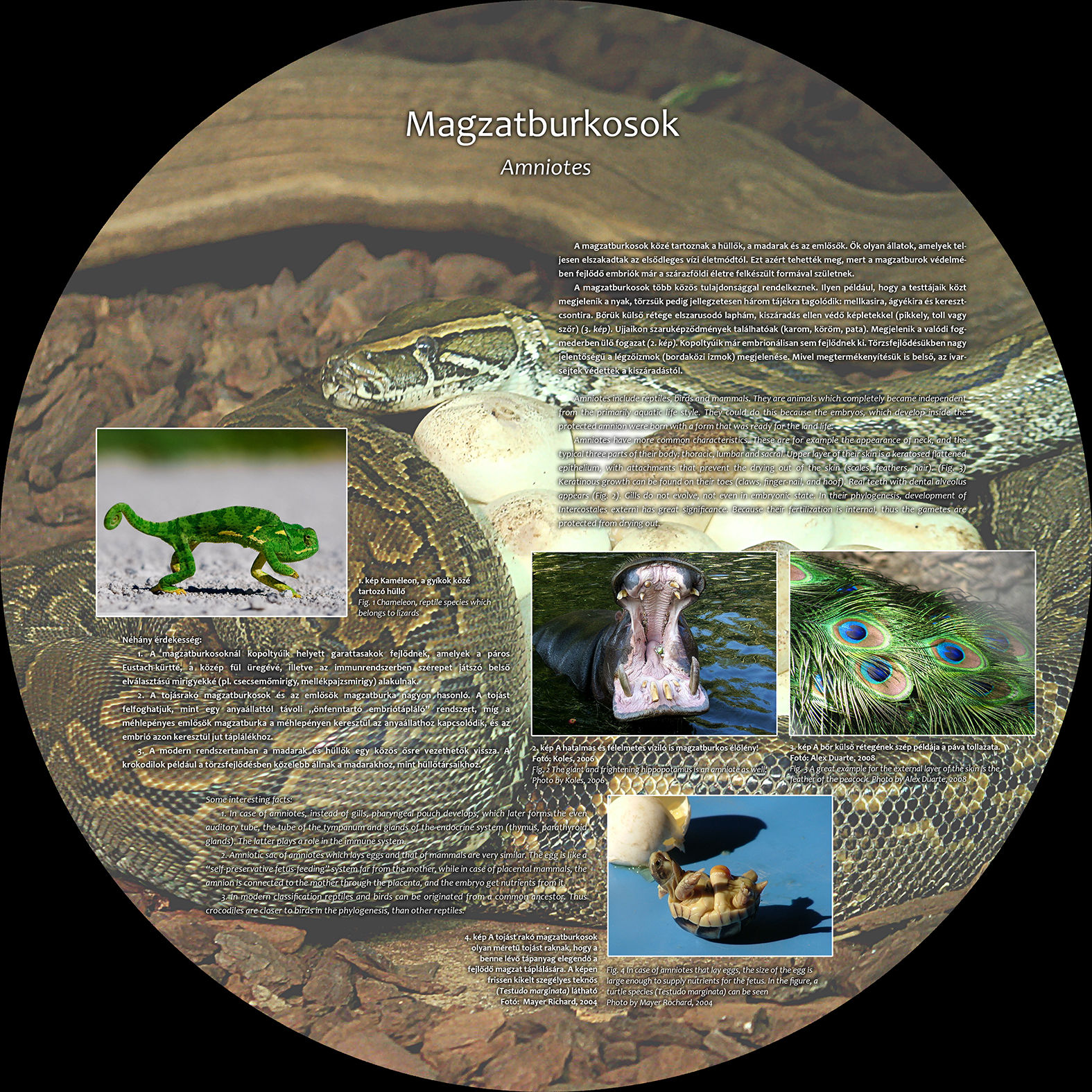
Fig. 2 The giant and frightening hippopotamus is an amniote as well! Photo by Koles, 2006
Fig. 3 A great example for the external layer of the skin is the feather of the peacock. Photo by Alex Duarte, 2008
Fig. 4 In case of amniotes that lay eggs, the size of the egg is large enough to supply nutrients for the fetus. In the figure, a turtle species (Testudo marginata) can be seen. Photo by Mayer Rochard, 2004